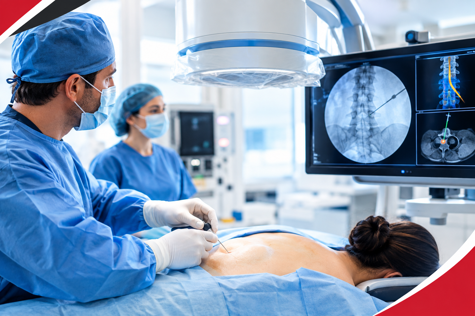
Pain procedures used to depend heavily on anatomical landmarks, clinical experience, and the physician’s ability to estimate where a needle should go.
Modern interventional pain management can be much more precise.
With fluoroscopy, ultrasound, and CT guidance, physicians can visualize relevant anatomy during selected procedures and guide a needle or treatment device toward a specific target.
Instead of relying entirely on what can be felt from the outside, the clinician can use imaging to understand the structures beneath the skin as the procedure is performed.
That shift matters because chronic pain is rarely just about where it hurts.
A patient may feel pain in the lower back while the relevant structure is a spinal joint, nerve root, sacroiliac joint, or another anatomical source. The location of pain can provide clues, but it does not necessarily identify the structure responsible for generating it.
Image-guided procedures are one part of a larger effort to make pain management Fayetteville NC service more targeted, deliberate, and anatomically informed.
What Image Guidance Actually Adds to a Pain Procedure
The phrase “image-guided” can sound more complicated than it is.
At its core, the idea is straightforward: the physician uses medical imaging to help identify anatomy and guide the procedure toward the intended target.
Different imaging technologies provide different types of information.
1. Fluoroscopy: Real-Time X-Ray Guidance
Fluoroscopy uses continuous or intermittent X-ray imaging to produce moving images during a procedure.
It is particularly useful when the physician needs to identify bony structures, such as parts of the spine, and confirm needle positioning during certain spinal injections and other interventions.
Fluoroscopy has been widely incorporated into interventional pain practice and remains an important guidance method for many spinal procedures.
One advantage is that the physician can obtain multiple views of the anatomy and confirm the position of the needle relative to the intended target.
Because fluoroscopy uses radiation, the amount of imaging should be appropriate for the procedure. Modern systems can also document fluoroscopy time and radiation dose as part of procedural records.
2. Ultrasound: Seeing Soft Tissue in Real Time
Ultrasound works differently.
Rather than using ionizing radiation, it uses sound waves to produce real-time images.
That can allow the physician to visualize soft tissues, nerves, blood vessels, muscles, tendons, and other structures that may not be adequately characterized by conventional X-ray imaging.
Ultrasound can also show the needle as it moves toward the intended target.
This makes it particularly useful for selected peripheral nerve, musculoskeletal, and soft-tissue procedures. Research comparing ultrasound with other guidance techniques continues to expand, although the advantages vary depending on the procedure and anatomical region.
3. CT Guidance: Detailed Cross-Sectional Anatomy
Computed tomography provides highly detailed cross-sectional images.
CT can be useful when complex anatomy requires a more detailed view than conventional fluoroscopy or ultrasound can provide. However, CT-guided pain procedures require specialized equipment and are not necessary for every intervention.
The choice of imaging technology depends on the anatomy being treated, the procedure being performed, the patient’s circumstances, and the physician’s clinical judgment.
There is no single imaging modality that is universally appropriate for every pain procedure.
Image Guidance Is Not a Guarantee of Better Pain Relief
It is tempting to assume that if a procedure is more precisely targeted, it must automatically produce better long-term pain relief.
The evidence is more nuanced.
A systematic review and meta-analysis comparing ultrasound- and fluoroscopy-guided interventions for back pain found no significant difference in post-procedure pain intensity or complications between the two approaches in the studies analyzed.
Functional outcomes showed a trend favouring fluoroscopy, although the difference was borderline.
Another meta-analysis examining ultrasound versus fluoroscopy for epidural injections in radiculopathy found comparable pain and functional outcomes in the studied groups, while reporting a lower risk of inadvertent vascular puncture with ultrasound-guided techniques in that analysis.
The diagnosis, patient selection, procedure choice, technique, and underlying condition still matter.
From “Where Does It Hurt?” to “What Structure Is Involved?”
Modern pain medicine increasingly works from a more detailed clinical picture.
Pain location is important, but it is only one piece of the assessment.
A patient with back pain may have symptoms related to different anatomical structures. Neck pain may involve cervical joints, discs, nerves, or surrounding soft tissues. Hip, shoulder, knee, and pelvic pain can similarly arise from several possible sources.
Before an intervention, the clinician may consider:
- The location and pattern of pain
- Physical examination findings
- Neurological symptoms
- Relevant imaging studies
- Duration and severity of symptoms
- Previous treatments
- Functional limitations
- The suspected pain generator
- Whether an intervention is appropriate at all
Image guidance then becomes part of the procedure itself rather than a substitute for diagnosis.
That distinction is important because a perfectly placed injection is not useful if the wrong structure was selected as the treatment target.
Which Pain Procedures May Use Image Guidance?
Image guidance is used across a range of interventional procedures, depending on the patient’s condition and the clinician’s approach.
Examples may include:
- Epidural injections
- Facet-related procedures
- Medial branch blocks
- Radiofrequency procedures
- Peripheral nerve injections
- Joint injections
- Musculoskeletal injections
- Other targeted nerve or pain interventions
The appropriate imaging method varies.
Fluoroscopy may be particularly useful when bony anatomy and spinal positioning need to be visualized.
Ultrasound can be valuable when the procedure involves superficial structures, nerves, blood vessels, or soft tissues. CT may be considered when detailed cross-sectional anatomy is important.
The goal is not to use the most technologically sophisticated option simply because it exists.
The goal is to use the appropriate tool for the anatomy and procedure.
The Role of Ultrasound Is Expanding
One of the more interesting developments in pain medicine is the growing use of ultrasound.
Ultrasound can provide real-time visualization without ionizing radiation and can show structures that are difficult to appreciate with conventional X-ray imaging.
A 2025 review described the increasing transition toward ultrasound-guided pain interventions and highlighted its ability to provide real-time anatomical visualization.
But this does not mean fluoroscopy is becoming obsolete.
For many spinal interventions, fluoroscopy remains highly useful. The two technologies are better understood as different tools rather than competing technologies where one must replace the other.
The physician’s experience with a particular procedure also matters.
Precision Is Only One Part of Good Pain Management
Technology can improve what the physician sees.
It cannot determine the entire treatment plan.
A modern pain evaluation still needs to consider the patient’s diagnosis, goals, functional limitations, previous treatment response, medications, medical history, and the risks and potential benefits of an intervention.
This is especially important for chronic pain.
Chronic pain can involve changes in the nervous system, musculoskeletal problems, nerve irritation, previous injury, degenerative conditions, and multiple overlapping contributors. Treating one anatomical structure may be appropriate in some patients but insufficient in others.
That is why image-guided intervention is best viewed as one component of a broader pain-management strategy.
What Patients Should Ask Before an Image-Guided Procedure
Patients do not need to understand every technical detail of fluoroscopy or ultrasound before undergoing a procedure.
But they should understand the reason for the procedure.
Useful topics to discuss with the treating clinician include:
What structure is being targeted?
Knowing the intended target helps connect the procedure to the suspected source of pain.
Why is this procedure appropriate for my condition?
Different diagnoses respond differently to different interventions.
Which type of imaging guidance will be used?
Fluoroscopy, ultrasound, and CT provide different information and have different practical considerations.
What is the goal?
The goal might involve reducing pain, improving function, helping confirm a suspected pain generator, or creating an opportunity for another treatment.
What are the alternatives?
An injection or procedure may not be the only reasonable option.
What are the risks?
Every procedure has potential risks, and those depend on the intervention, anatomy, medications, and individual health factors.
These conversations are often more valuable than focusing solely on whether a procedure is “advanced.”
The Future of Image-Guided Pain Management
The next stage of interventional pain care is unlikely to be about replacing one imaging technology with another.
It is more likely to involve increasingly individualized use of imaging.
Better visualization can help clinicians understand anatomy. Improved equipment can make procedures more efficient. Research can clarify which techniques provide meaningful clinical benefits for specific conditions.
At the same time, evidence-based pain medicine requires something equally important: recognizing when technology does not change the outcome enough to justify a particular approach.
That balance matters.
The purpose of image guidance is not to make a procedure look more sophisticated. It is to help the clinician perform an appropriate intervention with a clearer understanding of the anatomy involved.
Frequently Asked Questions
Are image-guided pain injections more accurate?
Image guidance can improve the physician’s ability to visualize anatomy and guide a needle toward a planned target. However, accuracy and clinical benefit are not identical. Evidence comparing guidance methods shows that outcomes can vary by procedure and anatomical region.
Is ultrasound safer than fluoroscopy?
Neither technology should be described as universally safer. Ultrasound does not use ionizing radiation and can visualize certain soft tissues and blood vessels in real time. Fluoroscopy provides important visualization of bony anatomy and remains valuable for many procedures. The appropriate choice depends on the procedure and patient.
Does image-guided pain management eliminate the need for surgery?
Not necessarily. Image-guided procedures are generally used for selected pain conditions and may be part of nonsurgical management. Whether surgery is appropriate depends on the underlying diagnosis and the individual’s clinical situation.
How long does an image-guided procedure take?
The duration varies considerably depending on the procedure, anatomical location, preparation, and whether additional steps such as diagnostic testing or medication administration are involved.
Can image-guided procedures be used for nerve pain?
Some image-guided procedures are designed to target nerves or structures associated with nerve-related pain. Whether one is appropriate depends on the suspected cause and location of the nerve problem.
The Bottom Line
Modern pain management is becoming increasingly precise, but precision should never be confused with complexity for its own sake.
Fluoroscopy, ultrasound, and CT allow physicians to visualize anatomy during selected procedures and make more informed decisions about needle placement and treatment targets. The best imaging approach depends on the procedure, the anatomy, the patient’s condition, and the clinician’s expertise.
Most importantly, the image is only one part of the treatment.
A successful pain-management plan begins with understanding the source and pattern of pain, determining whether an intervention is appropriate, selecting the right target, and choosing a technique supported by evidence.
Personalized Interventional Pain Care at Neurology & Pain Management Center
At Neurology & Pain Management Center, pain care is built around more than simply treating a painful area.
We bring together expertise in neurology, pain medicine, and sleep medicine, allowing complex pain concerns to be evaluated from a broader neurological and functional perspective.
Dr. Nailesh Dave is triple board-certified in Neurology, Pain Medicine, and Sleep Medicine, with a practice philosophy centred on accurate evaluation, evidence-based treatment, personalized care, and helping patients improve everyday function and quality of life.
When an interventional procedure is appropriate, the focus is on matching the treatment approach to the patient’s diagnosis, symptoms, anatomy, and individual goals rather than using a one-size-fits-all strategy.
Contact Neurology & Pain Management Center to schedule an evaluation of pain management in Fayetteville NC, and discuss a treatment plan tailored to your condition.